M-Mode Ultrasound

M-MODE Ultrasound:
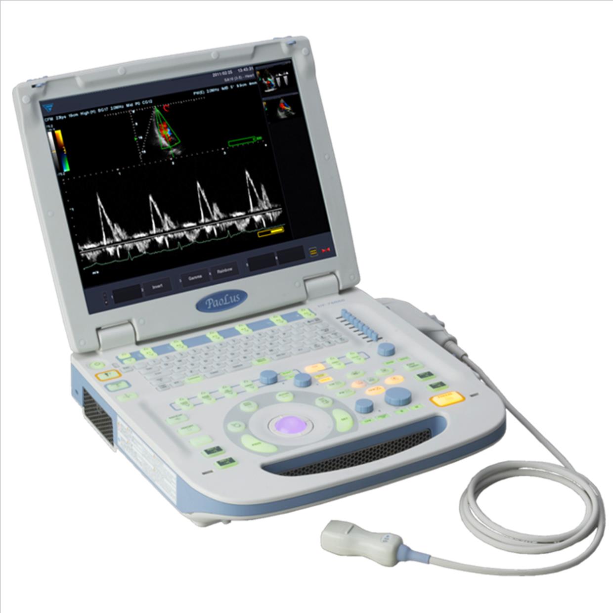
M-MODE Ultrasound is a method of echo cardio investigation using ultrasound imaging scanner
After B-Mode image information is obtained, by placing any reference line within the cross section, dynamic changes in time at that particulate place in terms of timing can be represented as a wave of changes in time, this is refereed as M-Mode ultrasound. Rate at which it is examined is called M-Mode speed.
M-mode echo-cardiograph is use to evaluate the morphology of heart structures, movement and velocity of cardiac valves and walls and timing of cardiac events. The ultrasound scanning positions for M-mode are mainly on the right thoracic wall. Two dimensional images produced in a parasternal long-axis and sort axis view are obtained and are used to produce an M-Mode trace. cross section of the left ventricle or right ventricle of the heart can be seen at various levels. At the papillary muscles, mitral valve and aortic valve. Measurements can then be made to determine, Fractional Shortening (FS) and anatomical measurements in one dimension in both systolic and diastole.
Angle free M mode
Anatomical M Mode or can provide easier line positioning and allow for related angle calculations corrections. Some times refereed as Anatomical M-Mode used within M mode echo cardio investigations and diagnosis of heart and functional dynamics of cardiovascular system in real time using an ultrasound scanner.

